On February 15, 2018 (the day after Valentine’s, no less) my female Dendrobates tinctorius ‘Robertus’ dart frog laid her first clutch of eggs. Of the 7 eggs, only 2 were viable after the first 24 hours. I thought it would be fun to take high-magnification microscopy images of their development over time and track the progress on this post. Throughout, I’ll share some tidbits of anuran developmental and natural history, geared especially for folks coming from the hobby world who might not have had the chance to study amphibian biology in depth. I am planning to update this post every few days with new progress shots, so be sure to check back in.
First, here is the happy mother:
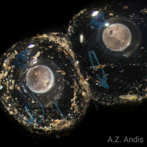
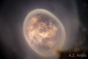
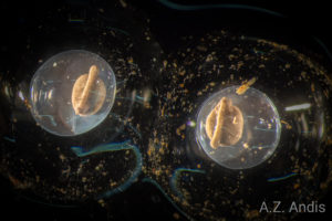
Day 1:
Here are the first images of the viable eggs, approximately 36-hours after oviposition and Gosner stage 12. At this stage, the embryo has gone through many cell divisions, but if you look closely, you can still make out the individual cells. For the first period of embryonic development, the zygote (fertilized, single-cell eggs) divides into a mass of multiple cells. Around the developmental period in these photos, the cell are beginning to migrate in order to form specific structures in the embryo. The dark indent on the surface, called the blastopore, is where the outer cells are closing in on themselves, like a deflating basketball that is being inverted. As the cells migrate inward, they create cavities that will form the body cavities of the fully formed animal. The blastopore will eventually become the anus, and the inwardly migrating cells will form the lining of the digestive track.
I also thought it might also be interesting to take a look at some of the non-viable eggs that were beginning to decompose. 
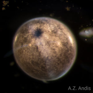
Day 3:
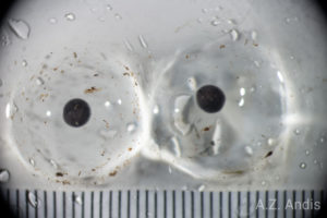
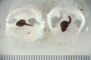
Approximately 72 hours, Gosner stage 12. The eggs are still developing. I took a scale shot to give a sense of their size. They measure 3.034 mm (L) and 3.180 mm (R) in diameter, which I estimate makes them about 14.6 (L) and 16.8 (R) cubic mm in volume. It’s worth remembering that, at this point, the embryo is still the same size as when it was a single-cell zygote after initial fertilization, even though it is now comprised of thousands of cells. The cells are dividing, but not growing. In the high magnification photos you can see that the blastopore indent is deforming and elongating. Inside the embryo, cells are beginning to form the notochord which will eventually become the nervous system. Although it doesn’t look like much, most of the structural components of the major life systems are already in place.
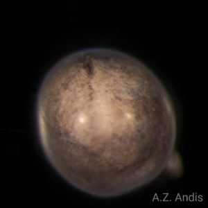
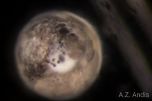
Day 5:
The embryos have elongated, measuring are 4.48 mm and 4.54 mm in length, almost 50% longer than yesterday, but the total mass should be about the same.
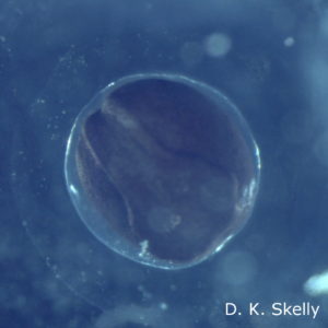
Gosner stage 18. The embryos developed a lot in just the last 48 hours. The embryos blew right through the end of gastrulation and into neurulation before I got these photos. So unfortunately, we missed the development of the neural plate, neural folding, and embryo elongation. As an overview, in the last photos the embryos were at the end of gastrulation. The cells had differentiated into cell layers, dividing the embryo into the ecotoderm (outer cell layer, eventually forming the skin, teeth, and most of the nervous system), mesoderm (middle cell layer, eventually forming most of the skeleton, internal organs, and muscles), and the endoderm (inner cell layer, eventually forming the lining of internal body cavities like the digestive track and lungs). So, at the end of gastrulation, the future fate of all cells were set, but the embryos still looked just like a ball of cells. Neurulation follows gastrulation, and as the name suggests, is characterized by the development of the proto neural system. These neural cells begin to migrate outward and develop ridges that will fold in on themselves during creation of the neural tube (which will eventually form the brain and spinal cord). (Since I didn’t capture a photo of the tinctorius eggs at this stage, I’ve include an example of a wood frog embryo at this stage as an example.)
As neurulation continues, the embryo starts to elongate and look more like a tadpole. By the later stages of neurulation (as in the photos today) most of the organ systems are fully established.
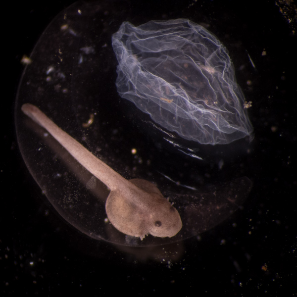
You can also see the very large yolk. Unlike bird, mammal, and reptile embryos, amphibian yolks form internally as part of the digestive track. Dendrobates eggs have much larger yolks than other frogs due to their particular larval ecology. In the wild, tinctorius eggs are deposited terrestrially, but the larvae are aquatic after hatching and require water immediately. So, these frogs have evolved a parental care strategy wherein the male attends the eggs, and upon hatching, transports them to a larger pool. The rearing pool can be hundreds of meters away from the egg site, and may take multiple days of transport. The large yolk ensures that the tadpoles will be hardy enough for the trip. Being hardy also helps the tadpoles once they are in the rearing pool since tinctorius larvae are cannibalistic.
You’ll also notice that the membrane around the embryo has expanded and is more pronounced. This called the vitelline membrane and it is analogous to the membrane surrounding the yolk in a chicken egg, the one that keeps a sunny-side-up egg from becoming a scrambled egg. Since tincortius eggs are laid out of the water, this membrane (along with the jelly layer) helps prevent drying.
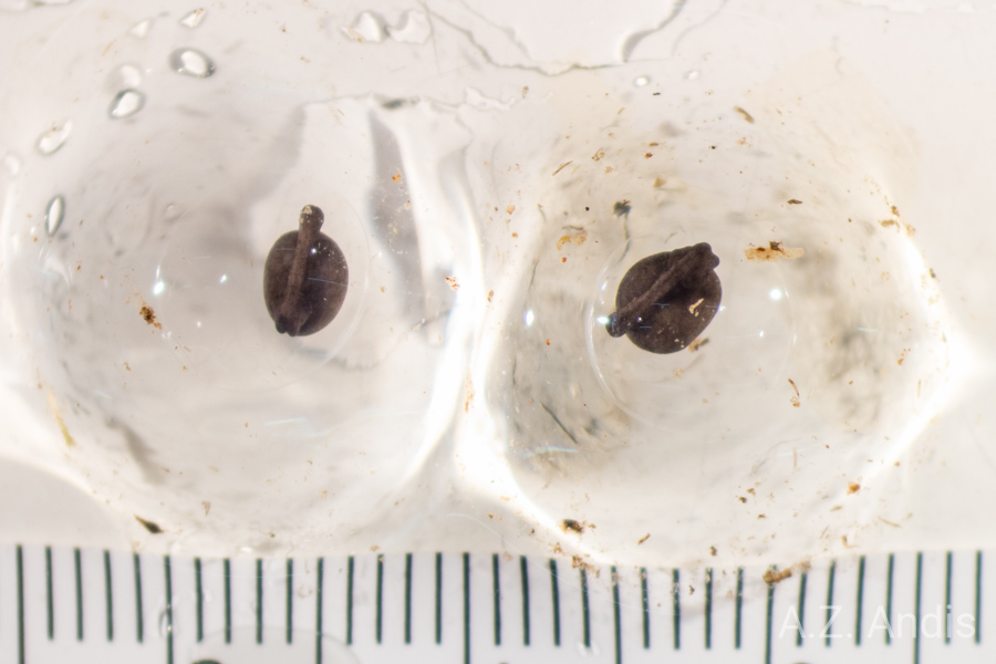
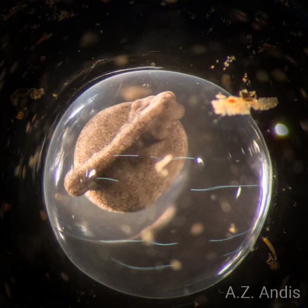
Day 6:

Gosner stage 19. We have gill buds! Those little nubbin-wings on either side are the sprouts of tiny gills. Up to this point, the cells of the embryo have been able to diffuse oxygen without special structures. But as the cells proliferate and some cells are buried deep within the developing embryo, simple diffusion can’t cut it. Over the next two or three days, the gills and circulatory system will take form and start pumping oxygenated fluid throughout.
Day 7:
Gosner stage 20. The gills are elongating and the simple heart, which is basically just a tube at this point, is pumping blood cells through the limited circulatory system.
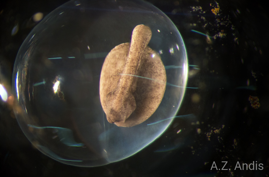
Day 8:
Gosner stage 21. The gills have formed almost to their external extent.
Tinctorius eggs are laid in oxygen poor conditions compared to many other frogs. As such, they have developed extremely long and filamentous gills compared to the “average” anuran. The greater the surface area of the gills, the more time there is for Co2 and O2 to diffuse across the lining of the gills to the jelly and, after hatching, to the water. If you look closely in the video, you will see the rhythmic expansion and contraction of the embryo. That’s the heart pumping. In the last portion of the video, you should be able to see individual blood cells flowing through the gills in time with the heartbeat.
Also notable at this point is that the nervous system is developing quickly, both the brain itself and the sensory organs. The proto-eye, while not externally visible, is developing as an outgrowth from the brain.
Stay tuned! The embryos are developing quickly and could hatch any time in the next few days!
Day 9:
The gills continue to branch and elongate. By this point, I think the heart has developed into two chambers, a huge architectural achievement from its origin as a single tube.
A caveat: it is worth mentioning at this point that my descriptions of developmental timing of internal organs are based on “average” anuran development. Not a lot of research has been conducted on dendrobatids, so most of these descriptions reflect ranid development. This is most clear in my attempts to assign Gosner stages to the embryos, as the ranid timing doesn’t sync entirely. Where research has been carried out, I try to give the specific description.
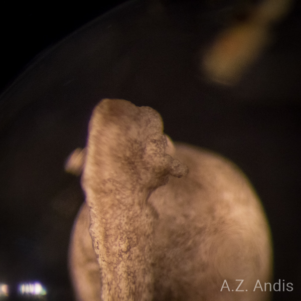
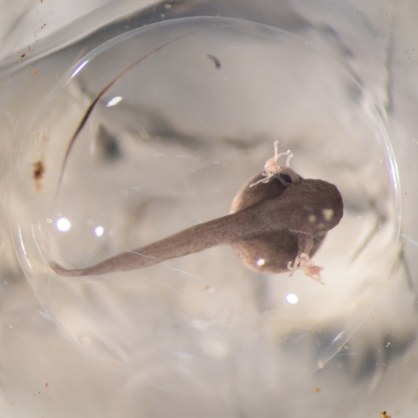
Day 10:
The gills are still growing, but should be reaching about their maximum extent. Soon, the gills will begin to atrophy and the animal will switch to internal respiration.
The eyes are just beginning to appear.
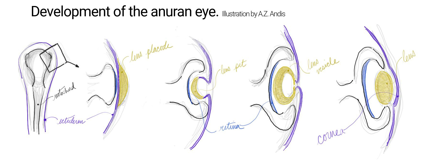
The embryonic eye forms as an extension of the developing nervous system that grows outward from the neural tube. As it reaches the surface of the embryo, the optic stalk differentiates into the component cell types of the eye. At this stage, the tip of the optic stalk at the skin surface is forming into a cup-like shape. In the coming stages, that cup deepens and widens. At the same time, the lens begins to form across the “rim” of the cup from the ectoderm. As the eye progresses, the lens becomes more “lens shaped” and the skin covering the eye becomes the cornea. If you are interested in learning more about the development of the anuran eye, I would suggest checking out Thomas Reh’s lab webpage.
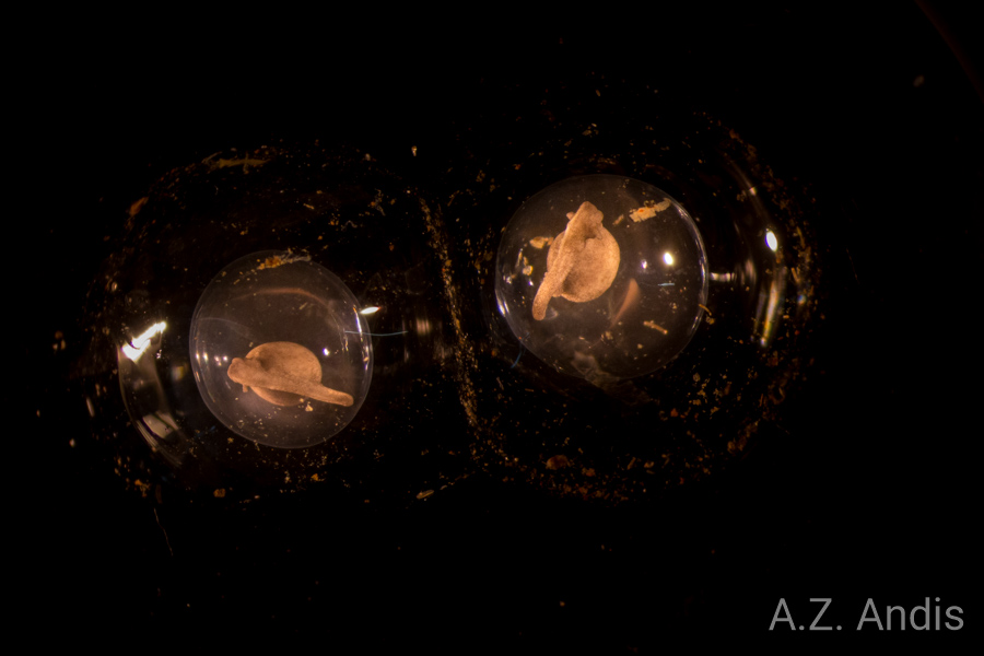
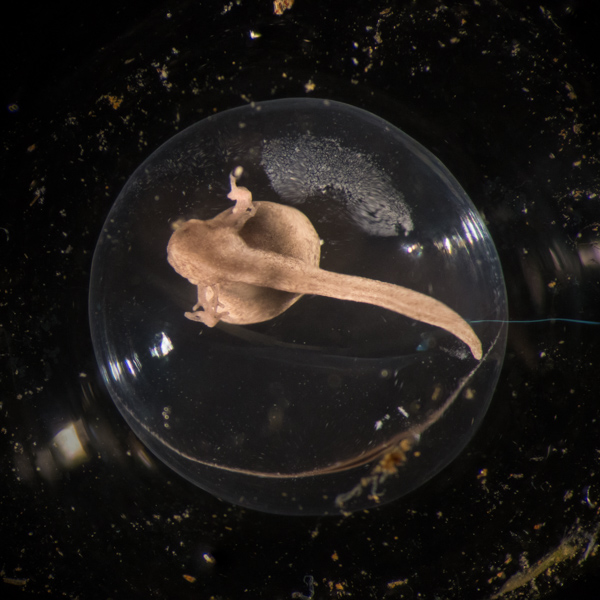
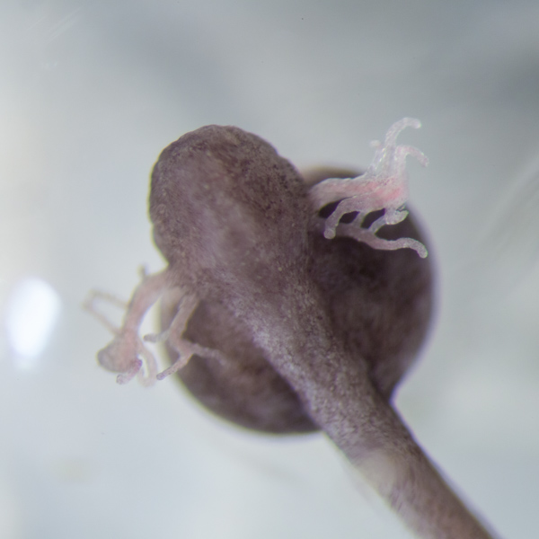
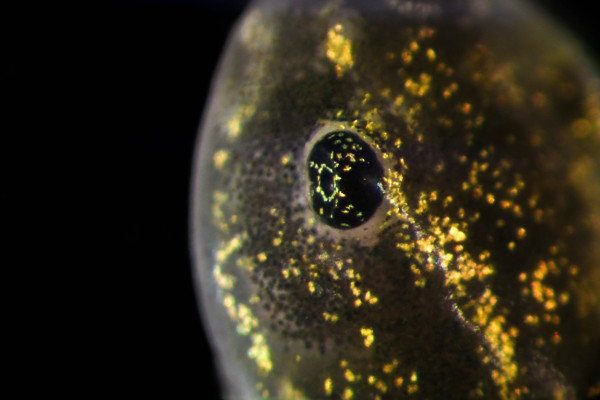
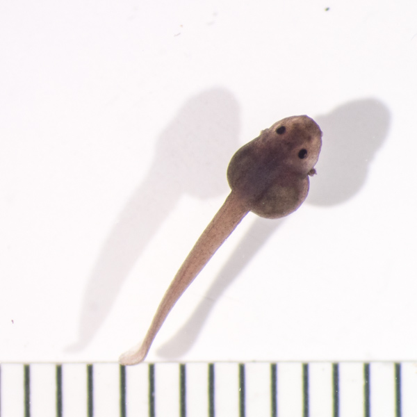
Day 11:
Today the eyes are well-along in development and obvious.
I outlined the formation of the eye yesterday, but I thought it would be good to also include a diagram of the formation.
To give you a sense of where the embryos’ eyes are heading, below is a close photo of a fully formed larval wood frog eye.
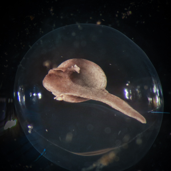
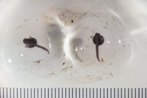
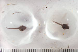
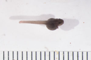
Day 12:
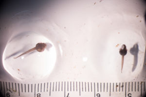
One of the embryos hatched today! The vitelline membrane is ruptured; however, the hatchling remains in the jelly and has not fully liberated itself. In the photo above, the animal on the right is the hatchling.

You’ll notice that the gills have begun to atrophy and shrink. As the heart becomes more efficient and the circulatory system grows, the distribution of oxygen to the body systems requires less surface area for gas exchange with the water. Also around this time, the primitive red blood cells of the early embryo transition into more efficient larval type red blood cells. Interestingly, there are a total of four generations of blood cells from embryo to adult. The cells change in shape, size, and even blood-type over the course of development. Even the organs producing blood cells shifts from kidneys, to the liver, and on to the spleen and bone marrow in adults.
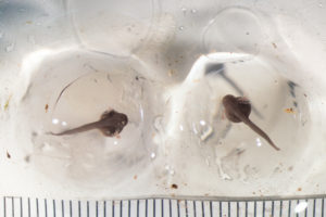
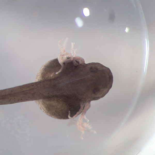
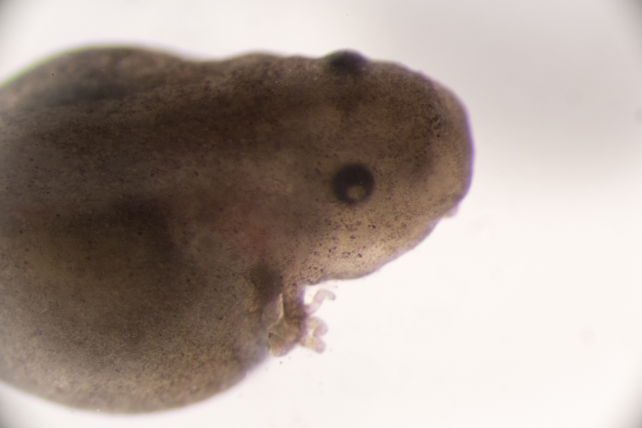
Day 13:
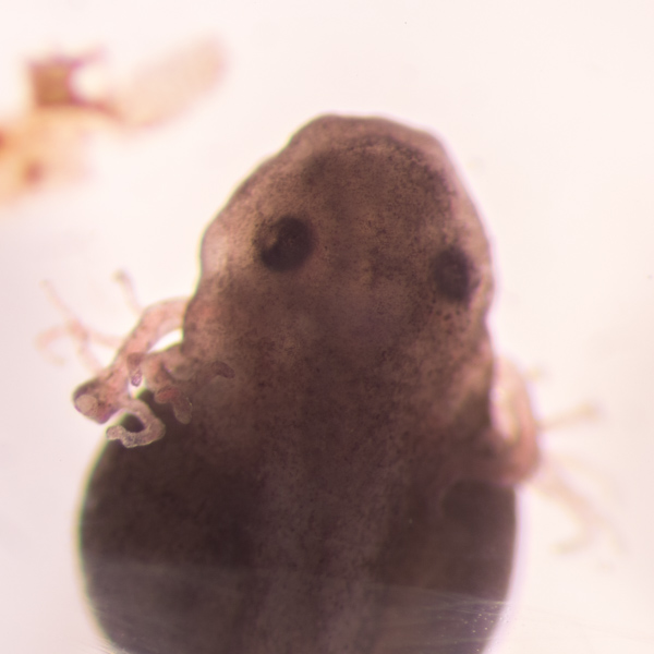
Both of the embryos have hatched out of the vitelline membrane, but remain in the egg jelly. They are very active and respond to movement and light. The gills have diminished quite a bit as the respiratory system switches to internal respiration. The eyes, while still covered by the outer skin layer, look much like a completed larval eye.
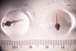
Day 14:
One of the hatchlings was deceased when I checked on it this morning. The tissue still looked pretty good; so I fixed it in a formalin solution to preserve its form.
The cornea is almost entirely transparent and the gills have almost complete atrophied. From this point on, the larva will assume the classic tadpole/pollywog body plan that is most familiar of anuran larvae.
References:
Hill, R. W., Wyse, G. A., and Anderson, M. (2016). Animal Physiology. 4 edition. Oxford University Press.
McDiarmid, R. W., and Altig, R. (2000). Tadpoles: the biology of anuran larvae. University of Chicago Press.
Rojas, B. (2014). Strange parental decisions: Fathers of the dyeing poison frog deposit their tadpoles in pools occupied by large cannibals. Behav. Ecol. Sociobiol. 68, 551–559.
Vences, M., Kosuch, J., Lötters, S., Widmer, A., Jungfer, K. H., Köhler, J., et al. (2000). Phylogeny and classification of poison frogs (Amphibia: dendrobatidae), based on mitochondrial 16S and 12S ribosomal RNA gene sequences. Mol. Phylogenet. Evol. 15, 34–40.
Vitt, L., Vitt, L., and Caldwell, J. (2013). Herpetology: an introductory biology of amphibians and reptiles. 4th ed. Academic Press.
Wells, K. D. (2010). The Ecology and Behavior of Amphibians. University of Chicago Press.
Weygoldt, P. (1987). Evolution of parental care in dart poison frogs (Amphibia: Anura: Dendrobatidae). J. Zoolog. Syst. Evol. Res. 25, 51–67.