Field-of-view:
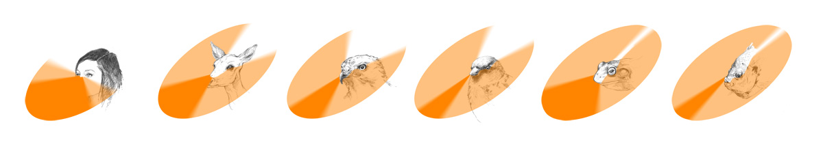
Images: Field-of-view for six taxa (human, deer, hawk, songbird, frog, and fish). The total arc of vision is represented in light orange while the binocular overlap is represented in solid, dark orange. Figure by A.Z. Andis with animal sketches by Bayla Arietta.
How an animal’s eyes are oriented largely determines its field-of-view and can tell you a lot about its ecology. Humans have forward-facing eyes which allows a large portion of the field-of-view to overlap and produce highly binocular vision across roughly 40 degrees of the entire 180 degrees of our field-of-view. The eyes of a bullfrog, on the other hand, are positioned near the top of the head and face outward. For a frog, this produces a 360 degree field-of-view in almost the entire visual arc but more limited binocular overlap.
In general, there is a trade-off between wide field-of-view and accurate depth perception. Since humans evolved to hunt and live in groups, good depth perception was more advantageous than a wide field of view. Frogs, on the other hand, faced selection pressure from pond-side predators, so detecting movement from any direction, even if fuzzy, was most advantageous.
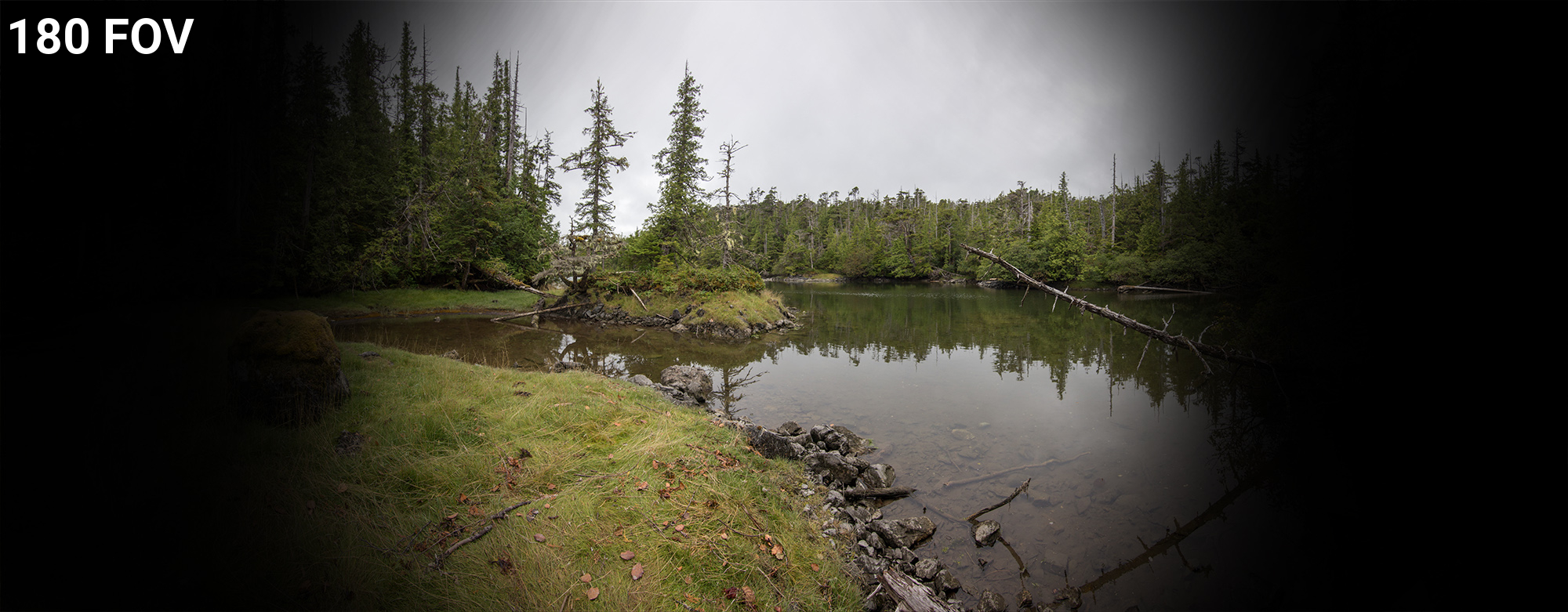
In this lesson, field-of-view is represented with a black vignette layer. For instance, in the slider below, a 180 degree field-of-view is compared to a 340 degree field-of-view.

Accommodation in Snakes
The ancestors of snakes were burrowing creatures that had no need for limbs or complex eyes. As such, these body elements were reduced or lost. As snakes adapted to occupy new habitats above ground, they re-developed complex eyes. Accommodation in snakes is achieved by the pushing the lens back in the eye with the muscles of the iris rather than deforming the shape of the lens itself.
Image: Fig. 1 from Caprette et al. 2004: Functional anatomy of lizard (A) and snake (B) eyes, illustrating major differences between the two general types. C, lizards focus by contracting large ciliary muscles (bm, cm) anchored to scleral ossicles (so) thereby applying pressure to the lateral surface of the lens (ln) via the annular pad (ap). D, snakes focus by moving their lens forward via increased pressure on the vitreous (vi) due to peripheral iris muscle (im) contraction. Abbreviations: an, anterior pad; bm, Brücke's ciliary muscle; cb, ciliary body; ch, choroid; cm, Crompton's ciliary muscle; cn, conus papilliaris; co, cornea; el, eye lid; fv, fovea; id, iris dilator muscle; is, iris sphincter muscle; ln, lens; re, retina; sc, scleral cartilage; sl, sclera; sp, spectacle; vi, vitreous; zf, zonular fibres.

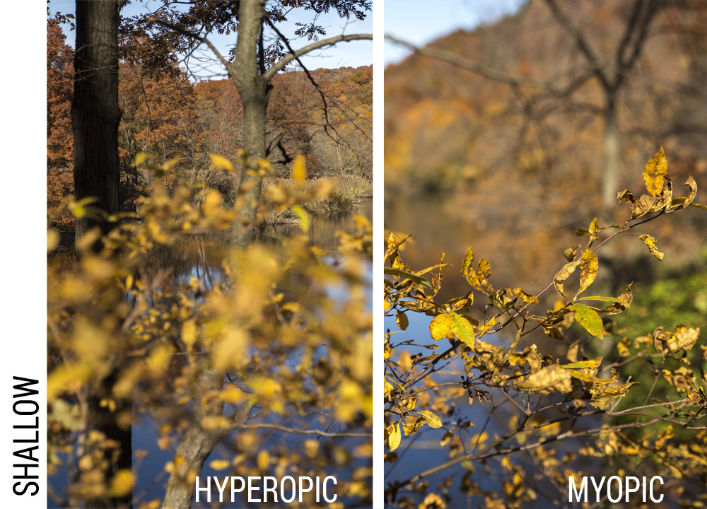
Focal field:
After passing through the lens, focused light falls on the retina which contains high densities of photoreceptor cells. Given plenty of light, the limiting factor that determines the sharpness of an image is how many photoreceptors can be packed into the retinal plane. In most species, high densities of photoreceptors are clustered near the middle of the retina to provide the sharpest image at the center of the field-of-view. This area of high-density reception is called the fovea.
In human eyes, the fovea is small and restricts the clearest vision to just the central 5-10 degrees of our field of view. This is why it is extremely difficult to read unless you look directly at a line of text.
As a test, lock your eye onto the "A" at the beginning of this sentence, then without letting your eye move, try to read the rest of the sentence. While you can see that the entire line of text falls within your vision, you probably had a lard time making out even one or two words past "As". Such a restricted field of clarity is no problem for humans since we can easily swivel our head and eyes, but is more of a consideration for animals with relatively fixed heads and eyes like snakes and frogs.
In this lesson, focal field is represented as a blurred vignette, like in the slider below.


Pupil Shapes
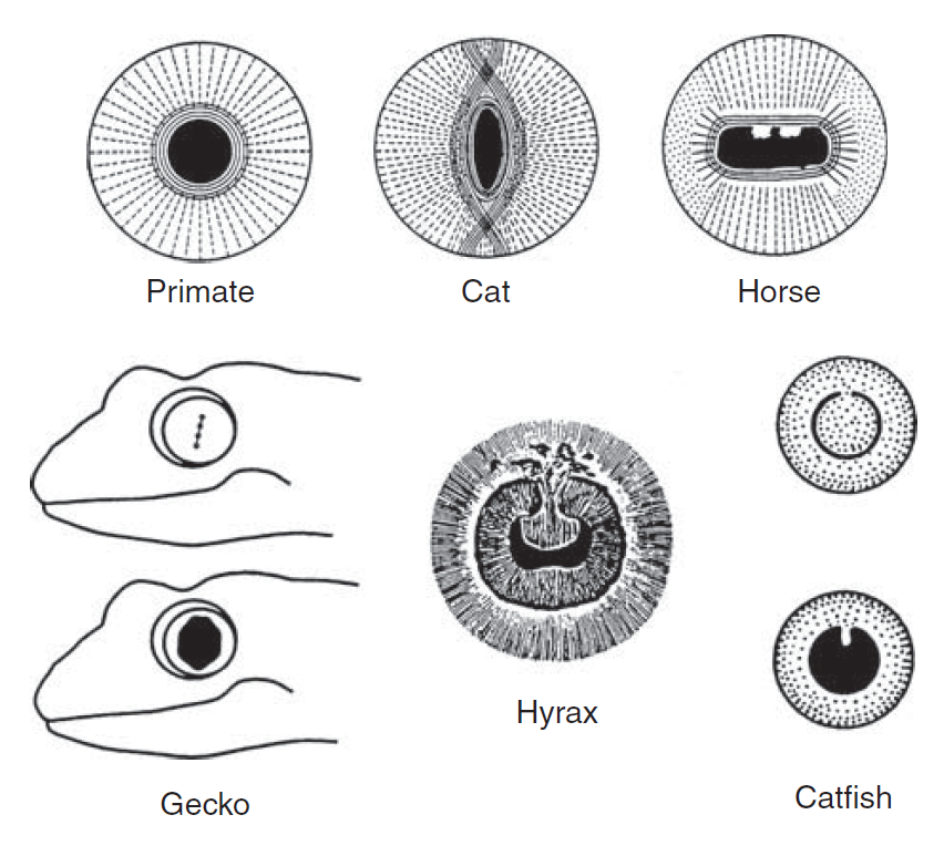
Vertebrate eyes have an iris--the ring of muscles that open and close around the eye opening to control the amount of light entering the eye. In humans and most vertebrates, the iris forms a round pupil. However, round irises cannot close tightly because the muscles bunch up. To prevent this, animals that must see in a range of light, like cats, geckos, and frogs, have slit-like openings that can be closed almost completely. Other vertebrates, like fish and snakes, have immovable irises and have adapted other mechanisms to manage the amount of light entering the eye.
Image: Fig. 5.11 from Land and Nilsson 2012: Pupil shapes in vertebrates. Top row: round and slit shaped pupils in mammals, showing how the cat’s slit pupil can close further than the circular primate pupil. Iris closer muscles are continuous lines and opener muscles dashed lines. Bottom row: gecko pupil contracts to four ‘pinholes’ in the light. The hyrax or coney ( Procavia, a small desert mammal) has a pupil partly closed by a central operculum, which acts as a sunshade. A similar mobile operculum is present in some fish, such as the catfish Plecostomus. Combined from Walls (1941).
Spectral sensitivity:
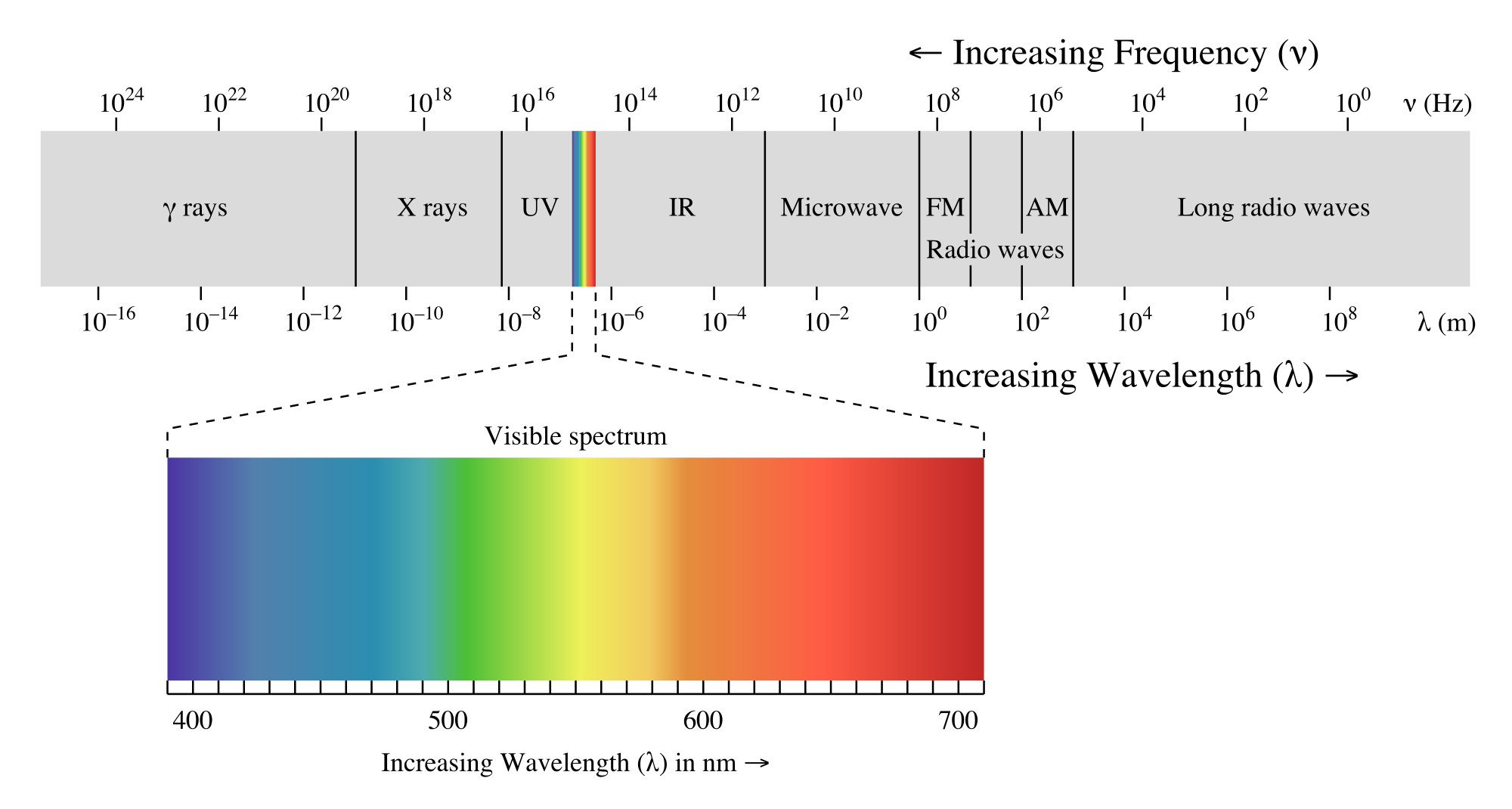
Image: Diagram of the electromagnetic spectrum, including human visible light. Figure by Philip Ronan, usage under Creative Commons.
Vertebrates have two types of photoreceptors: rods, that cannot discern color but can function in low light (although blurrily), and cones, that react to narrow spectral frequencies of light, and allow for color vision if two or more cone-types are present.
Photoreceptors respond to different frequencies of light depending on the pigment they contain. There are five types of pigments employed in vertebrate eyes, four color cone pigments and the pigment in rods which is sensitive to blue-green wavelengths. Cone pigments are sensitive to long, red wavelengths; medium, green wavelengths; short, blue wavelengths; or super-short, violet and ultraviolet wavelengths.

Image: A rudbeckia flower as seen in the human visible spectrum (left) and in ultra-violet frequencies (right). Notice that patterns visible in UV light are not apparent in other frequencies. Photos by A.Z. Andis.
It should be no surprise that animals have evolved to see some wavelengths of light better than others. The sun is the primary source of light energy for all animals. We humans are most intimate with the relatively narrow band of the electromagnetic spectrum from the sun between about 380 to 760 nm. These wavelengths encompasses our visual range across the rainbow from violet to red. Other species have evolved to see shorter wavelengths, in ultraviolet, and longer wavelengths, in infrared. Other species only see limited portions of the human visible spectrum. So, why did evolution result in the restricted frequencies of some species? And why are all species more or less restricted to such a narrow sliver of the entire electromagnetic spectrum?
Shorter wavelengths in the blue and ultraviolet spectrum are easily absorbed and scattered by water and air particles (that is why sunsets turn red and orange as the light travels through more atmosphere at the horizon). At the longer end of the spectrum, infrared light tends to dissipate too quickly and does not have enough energy to be useful for sight, especially over distance. Thus, due simply to physics, most animal vision is tuned within the Goldilocks zone from infrared to ultraviolet.
Within the Goldilocks frequency range, evolution has tuned animals into wavelengths most useful for their ecological situation. In water, ultraviolet light only penetrates about 50 meters, making it useful to species at shallow depths but useless for deep-swimming species. Similarly, yellow and red light cannot penetrate water far beyond 50-100 meters. So, fish tend to have evolved the highest sensitivity to the light available at their preferred depth.
In addition, the high energy of short wavelengths can be damaging to cells (which is why we wear sunscreen at the beach to block UV). In many cases, animals that are active during the daytime have evolved ways to shelter their cells from these frequencies rather than absorb them.
It is difficult to pack lots of color cones into the retina. Color cones are not usually distributed evenly across the retina. Many animals, including humans, pack most of the color-sensitive cones in the center of the field-of-view and sacrifice color vision around the periphery. In this lesson, the effective cone density is represented with a desaturation vignette, as demonstrated in the slider below.