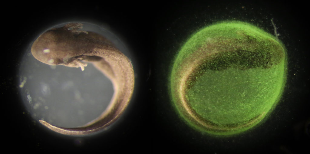
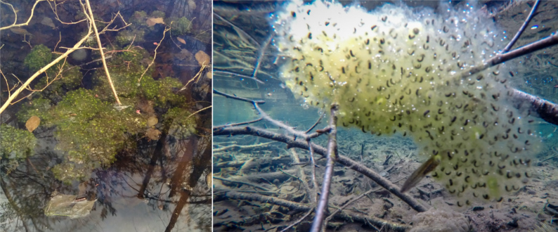
As part of my dissertation research, I spent the spring monitoring the development of wood frog eggs. Some of the eggs I brought to the lab, removed from the jelly coat and reared in incubators. I left the rest of the eggs in the ponds and checked on them daily until they hatched. Although not part of my research, I thought it would be interesting to take photos comparing the wild eggs and algae symbionts to the lab-reared eggs without. The relationship between this alga species and only a handful of amphibians is one of the most unique stories in biology. To the best of my ability, I’ve chronicled the Green Egg symbiosis story below.

Green Eggs
Folks have been fascinated by the coincidence of algae and amphibian embryos for a long time. One of the first notes was published in 1888. Orr was studying amphibian skeletal and nervous system development. He needed embryos and had collected egg masses of a few local species. He was surprised to see that the salamander eggs he procured seemed “to present a remarkable case of symbiosis.” He noticed that there were algae cells situated inside of the egg membrane.
This is surprising because that membrane, called the vitelline membrane, is impermeable to everything but ions and small molecules, which certainly excludes entire algae cells. So the only other option is that the algae entered the embryo before the vitelline membrane had formed. But this would be no less surprising because, like basically all animals, the vitelline membrane is formed in the ovaries, and in amphibians, extra layers of jelly are added in the oviducts, long before the egg is exposed to pond water.
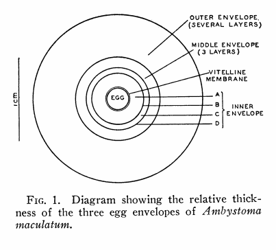
In the figure above (from Gilbert 1942) depicting the layers surrounding a salamander egg, it is clear to see that there are many layers acting as barriers between the pond water and the egg.
Which came first–the algae or the egg?
So, Orr was left with the plaguing question: Which came first—the algae or the egg? Perplexed, he wrote, “I have not discovered how the Algae enter the membrane, nor what physiological effect they have on the respiration of the embryo, but it seems probable that in the latter respect they may have an important influence.”
It turns out that Orr was right, algae do have an important influence on the embryos, but it was not demonstrated until the 1940s. The phenomenon continued to perplex scientists for decades. By the 1940s, algae had been reported in the eggs of multiple salamander species and also wood frogs across the continent from California to New England and Virginia. Gilbert (1942) was the first to explore how the algae came to enter the egg and also studied the importance of the algae-egg relationship.
As to the entrance of the algae into the egg, he first assumed that the algae were present in the ovitract of the female salamander. He washed the oviducts of salamanders and tried to culture the solution, but no algae developed. If algae were not present in the ovitract, then they enter from the pond after the eggs are deposited. To confirm, Gilbert took a recently inseminated female and allowed her to lay eggs in an aquarium with pond water then removed her and allowed her to lay another clutch in an aquarium with algae-free tap water. Algal cells were present in the eggs laid in pond water within hours, but no algae grew within the tap water eggs after a few days. At the end of this experiment, Gilbert put the eggs from the tap water aquarium in a natural pond. Within a few days, algae had penetrated the jelly membrane.
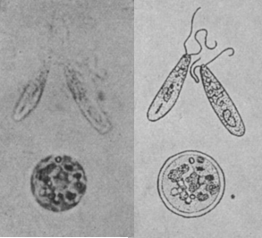
By closely studying the eggs, he found that the algae transformed as they penetrated the egg. When free-swimming in the pond, the algal cells are long and oval with flagella used for locomotion. As the algae penetrate the egg layers, they grow in size, become spherical in shape, and lose their flagella. It is these large, non-mobile cell-types that rest on the inner membrane and create the “verdant blanket” (as Gilbert describes it; 1942, p. 220).
To demonstrate symbiosis, he compared hatching rates in egg masses kept in light and dark to prevent photosynthesis, finding that light and algae increased hatching success. Although algalogistis (yep, that’s a thing) hadn’t officially recognized the alga species, folks who worked with amphibian eggs called it Oophila amblystoma (“Oo” and “phila” come from the Greek words “egg” and “love,” and “amblystoma” is the genus name of the salamander species the algae cohabitates with. So, the name just means “an algae that likes salamander eggs”).
Just a few years ago, researchers took a closer look at the Oophila algae. They wondered if this was really one species or a clade of algal species, and if it the latter, did different algal species associate with specific amphibian species? To answer those questions, they sequenced the genome of algae from four species across the continent.
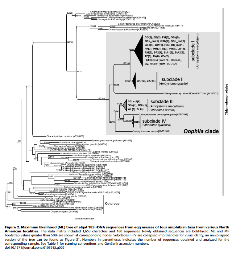
It turned out that the Oophila are closely related, close enough to be considered a single clade or species, but within the group there are subclades that tend to associate with each host. However, the researchers also found evidence that different subclades of the algae occasionally defect and switch to other host species.
Friends or just roommates?
Since Gilbert’s era, we’ve learned a lot about the economy of the biological transactions between the algae and the embryo. Using microelectrodes inserted within the egg membrane, Bachmann et al. (1986) measured oxygen concentration within the egg in contrast to the outside water in both light and dark. In the light, the algae worked overtime, producing so much oxygen through photosynthesis that the oxygen production exceeded that needed by the embryo and the environment inside of the egg became super-oxygenated, even when the surrounding water was anoxic (oxygen-poor).
This extra photosynthetic boost is more than just a little helpful. Most amphibian embryos get oxygen by diffusion across the egg membrane from surrounding water or air. Rinder and Friet (1994) wondered if diffusion of oxygen from the surrounding water alone could support amphibian species that had evolved with algal symbionts. They measured oxygen gradients across the cell membrane and injected dye in egg masses to see how much water would be able to flow past the eggs. In wood frogs, they found that the egg mass was loose enough and contained enough channels that water diffusion alone could support respiration. However, spotted salamander egg masses are dense and water does not penetrate to the interior eggs. In their case, the innermost eggs would suffocate without their personal algal oxygen factories. (Note: Hutchinson and Hammen (1958) inferred this relationship many decades before through experimentation rather than directly measuring O2 within the eggs.)
This plant-animal relationship is a multifaceted exchange. The reciprocity extends past simple O2 and CO2 cycling. The algae also benefit from the nitrogen waste produced by the embryo, which limits the ammonia buildup in the egg which is toxic to embryos (Goff and Stein 1978).
Friends or more-than-just-friends?
This symbiotic relationship is certainly interesting, but not altogether surprising. After all, lots of animals host symbiotic microbes, even humans house a panoply in our guts and all over our skin (Gilbert et al. 2018). These types of ectosymbionts, commensal organisms that live in and on other organims’ bodies but outside of the tissue are common.
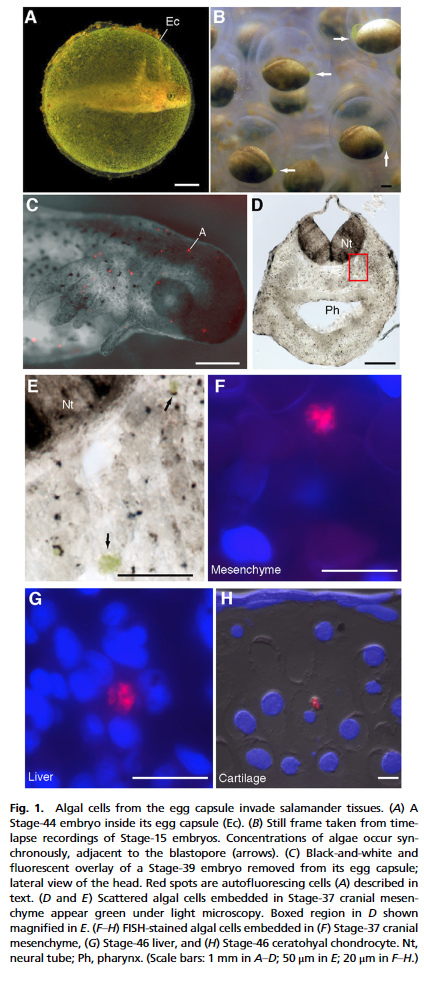
But the green egg story gets even more fascinating. In 2011, Kerney et al. used a technique that binds only to algae DNA and causes it to glow under fluorescent lights (fluorescent in situ hybridization (FISH)). In doing so, they could see algae cells inside(!) of the salamander tissue and cells. This makes this salamander-algae relationship the only example of endosymbiosis in a vertebrate ever seen! Previously, this type of animal-plant union was thought to only be possible in simpler organisms like coral and mollusks.
Friends or frenemies?
So, not only do these free-swimming algae manage to invade the egg membrane, but they even make it all the way inside of the embryonic cells. To some extent, this makes sense for the algae. Why would you want to live out in the pond water with variable temperatures and predators when you could live inside a nice cozy egg with abundant CO2 and nutrients? But once inside the egg, why would algae invade inside an animal cell where there is less access to light for photosynthesis?
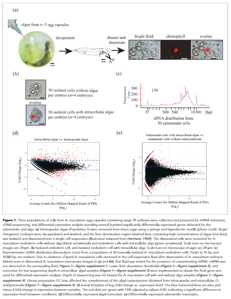
A team of researchers (Burns et al. 2017), including Kerney, wanted to figure out the riddle. They isolated and sequenced the genes expressed by salamander cells with and without algae cells from inside and outside of the animal cell. Inside the animal cells, algae seemed to be under more stress, to the point that they switch from photosynthesis to fermentative energy production, something that would normally only happen in highly unfavorable environments. On the other hand, salamander cells with algae were unfazed and even seemed to suppress their immune response which would ordinarily attack foreign cells, perhaps to keep the algae within. For algae, then, there appears to be a risk-benefit balance that drives them inside of the egg where conditions are favorable, but risk being captured inside of the embryonic animal cells.
Better than Seuss
After over a century of investigation, the story of the Green Eggs has gotten more and more interesting with finer and finer resolution. The saga goes to show how the naturalist’s intrigue can inspire a cascade of illuminating research.
Understanding this complex relationship can also help us understand threats to amphibian populations. Herbicides can kill the symbiotic algae and result in lower hatching success and reduced developmental rates. Olivier and Moon (2009) tested the effect of the herbicide atrazine on spotted salamander egg masses. They found that even low concentrations of the chemical killed the algae. In addition to the direct effects of the chemical, the loss of the symbiont had negative repercussions.
References:
Bachmann, M. D., Carlton, R. G., Burkholder, J. M., and Wetzel, R. G. (1986). Symbiosis between salamander eggs and green algae: microelectrode measurements inside eggs demonstrate effect of photosynthesis on oxygen concentration. Can. J. Zool. 64, 1586–1588.
Burns, J. A., Zhang, H., Hill, E., Kim, E., and Kerney, R. (2017). Transcriptome analysis illuminates the nature of the intracellular interaction in a vertebrate-algal symbiosis. Elife 6. doi:10.7554/eLife.22054.
Gilbert, J. A., Blaser, M. J., Caporaso, J. G., Jansson, J. K., Lynch, S. V., and Knight, R. (2018). Current understanding of the human microbiome. Nat. Med. 24, 392–400.
Gilbert, P. W. (1942). Observations on the Eggs of Ambystoma Maculatum with Especial Reference to the Green Algae Found Within the Egg Envelopes. Ecology 23, 215–227.
Goff, L. J., and Stein, J. R. (1978). Ammonia: basis for algal symbiosis in salamander egg masses. Life Sci. 22, 1463–1468.
Hutchison, V. H., and Hammen, C. S. (1958). Oxygen utilization in the symbiosis of embryos of the salamander, Ambystoma maculatum and the alga, Oophila amblystomatis. Biol. Bull. 115, 483–489.
Kim, E., Lin, Y., Kerney, R., Blumenberg, L., and Bishop, C. (2014). Phylogenetic Analysis of Algal Symbionts Associated with Four North American Amphibian Egg Masses. PLoS One 9, e108915.
Olivier, H. M., and Moon, B. R. (2010). The effects of atrazine on spotted salamander embryos and their symbiotic alga. Ecotoxicology 19, 654–661.
Orr, H. (1888). Memoirs: Note on the Development of Amphibians, chiefly concerning the Central Nervous System; with Additional Observations on the Hypophysis, Mouth, and the Appendages and Skeleton of the Head. J. Cell Sci. s2-29, 295–324.
Pinder, A., and Friet, S. (1994). Oxygen transport in egg masses of the amphibians Rana sylvatica and Ambystoma maculatum: convection, diffusion and oxygen production by algae. J. Exp. Biol. 197, 17–30.